
Bio-Magnetic Reflex Scanner: Theory, Results, Technique
Abstract:
OBJECTIVE: To determine a faster and more effective way to scan neurolymphatic reflexes. SUBJECTS: Fifty randomly selected existing patients were used in the experiment. DESIGN: Three neurolymphatic reflexes were scanned to see if an indicator muscle was inhibited by having the patient therapy localize each reflex and stressing these reflexes with south pole magnetic energy in combination with the colors red or blue to activate the sympathetic/parasympathetic nervous system. SETTING: Private office of Michael Lebowitz, D.C.. RESULTS: Many more positive results were discovered by exposing the patient to the south pole of a magnetic wand when the south pole was colored red or blue. CONCLUSIONS: By screening neurolymphatic reflexes with a red or blue colored south pole of a magnetic wand one can discover many cases of positive results that were initially missed by scanning neurolymphatic reflexes in the clear.
Key Indexing Words
Magnets, Sympathetic Nervous System, Parasympathetic Nervous System, Red, Blue, Neurolymphatic Reflex.
Introduction/Background
Screening for and treating organ dysfunction is a fundamental component of Applied Kinesiology (AK). Since the 1930’s neurolymphatic (NL) reflexes have been correlated with different types of dysfunction in various organs and glands. Thanks to Dr. George Goodheart these reflexes were introduced to applied kinesiology and have been utilized for this purpose with much success by applied kinesiologists for many years. One method utilized to assess organ “health” is by using an indicator muscle and having a patient therapy localize (TL) the corresponding NL points. When screening these reflexes, one wants to minimize the number of false negatives, allowing for an optimal diagnosis of the patient, and a correct treatment plan. Often times a reflex may show as functioning normally “in the clear”, but this may change as soon as the patient undergoes the slightest amount of stress to the corresponding organ. By inducing a sympathetic nervous system stressor to the reflexes being screened, non-optimal functioning organs may be detected, which tested normally in the clear. Although less common, the other extreme can also happen in a patient being “too calm and relaxed”, and if exposed to a stimulant of the parasympathetic nervous system an organ dysfunction can be detected.
All magnets contain a north and south pole. The North Pole of a magnet is also referred to as being “negative” and the South Pole is referred to as being “positive”. According to Dr. William Philpott the “negative field” is beneficial for the body while the “positive field” causes a state of stress and disease (1). The North Pole of a magnet also has been shown to have an alkaline affect on ones body, while the South Pole has an acidic affect. Dr. Philpott also noted how the south pole of a magnet increases inflammatory reactions and the growth of noxious microorganisms inside the body (2).
While the south pole of a magnet can be used to put the body in a sense of heightened awareness (3), one can also use color to stimulate the sympathetic or parasympathetic nervous systems. According to the
By using the south pole of a magnet one can heighten the body’s sense of awareness, and then stimulate the parasympathetic (using the color blue) or sympathetic (using the color red) nervous systems to scan for organ dysfunction. Using a facilitated muscle as an indicator muscle one can scan appropriate neurolymphatic points for organ dysfunction checking for inhibition of the indicator muscle when a red or blue colored south pole of a magnet is placed over the corresponding neurolymphatic point.
Materials and methods
We made a magnetic wand that has a south pole facing magnet on both ends. One end has a red color added to the end, while the other has a blue color (called a bio-magnetic reflex scanner). We decided to use three different anterior NL reflexes: those for the adrenals (bilaterally), pancreas, and reproductive organs (bilaterally). First we had the patient TL each reflex and recorded whether it caused inhibition of a strong indicator muscle. We then had a patient hold the south pole of a magnet wand over the NL reflexes, scanning for inhibition of an indicatory muscle. Thirdly we placed a red piece of construction paper in front of both eyes while the patient therapy localized the reflex and tested an indicator muscle and recorded the result. Then we applied the red/south pole of the device over the NL reflexes and checked if it inhibited an indicator muscle and recorded the result. We then did the same with blue as we did with the red.
Results
Fifty patients were screened using the above mentioned methods for NL reflexes weakening a strong indicator muscle. Having the patient TL the NL reflex showed many fewer positive results compared to a patient using the south pole of a magnet or looking at the color red while therapy localizing the corresponding reflexes.
Each NL reflex scanned for showed the greatest number of positive tests when a red colored south pole of a magnet was used to screen the NL reflex. In addition, several additional positive results were found when scanning with either looking at the color blue, or using a blue south pole of the magnetic wand. (Figures 1, 2, 3)
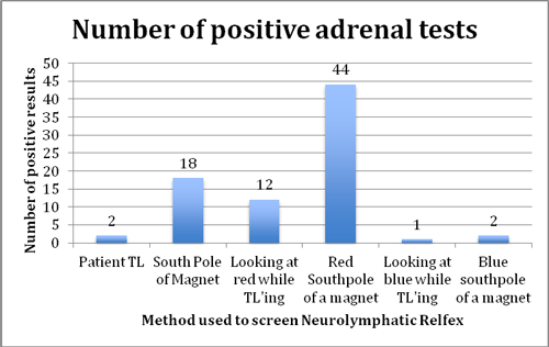
Figure 1. Adrenal NL reflex was checked and the results were recorded for six different methods of screening.
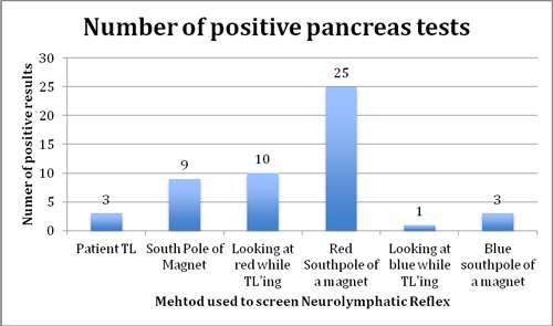
Figure 2. Pancreas NL reflex was checked and the results were recorded for six different methods of screening.
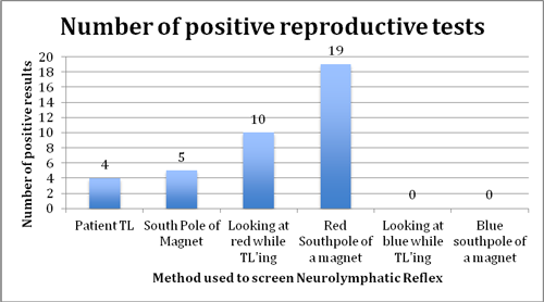
Figure 3. Reproductive NL reflex was checked and the results were recorded for six different methods of screening.
Discussion
The red/south pole combination by far showed the most positive tests with many unique findings that did not show in the other fashions. For instance, the NL reflex for the adrenals was positive 88% of the time with this method, as compared to only 4% with normal patient TL. This appears to confirm the fact, as noted by many previous AK researchers including this author- that applying the right stress can be very important to uncover relevant findings in the patient that can be helpful in enhancing his or her outcome. Of course, in these patients, we follow through with structural and nutritional support and retest these reflexes during future visits. Structural treatment involves rubbing the positive reflex while the reflex is exposed to the colored pole of the magnet that caused the inhibition response. Often times we need not treat these directly but correcting other core findings such as dysbiosis, toxic metals, food toxins, etc. as we have previously written about will bring resolution to these NL findings. In those cases we are using the tests to help monitor patient progress.
Over time we have also found the south pole/red combination has been a very useful way to find “hidden subluxations”, positive acupuncture points, etc.
Conclusion
Scanning NL reflexes using a magnetic wand with blue and red colored south poles is a fast and efficient way to scan for organ dysfunction. In addition to being able to easily scan the reflexes in an efficient manner, many false negatives were prevented, that were initially found with the traditional method of simply having the patient TL the various NL reflexes. Also by assessing if the organ is stressed by the parasympathetic vs sympathetic nervous system one can tailor the treatment plan in the attempt to either “increase” or “decrease” the functioning of the specific organ or gland.
This instrument is called the Bio-magnetic reflex scanner and is available from Mid American Marketing 1-800-922-1744
References:
1. "In Honor of Dr. William H. Philpott, M.D." True North Magnetics. 1 Jan. 2010.
Web. 30 Aug. 2011.
<http://www.truenorthmagnetics.com/pdf/True_North_Newsletter.pdf>.
2. Philpott, William H. MD "Magnetic Facilitated Central Nervous System
Biofeedback Response Test - Substance Response Test." Philpott Medical
Center (1990). Print.
3. Lebowitz, Michael D.C., “Biomagnetic Neurolymphatic Technique”, Proceedings of
the Winter Meetings of ICAK, Vol. 2, 1991-1992.
4. Ingersoll S. Syntonics as reading enhancement techniques at the Livingston
Developmental Academy (presented at 66th Annual Conference Light and Vision,
Vancouver, CN, 1998). Journal of Optometric Phototherapy 1999.
5. Silva, Amanda. "Color Therapy." Health Psychology Home Page. Vanderbilt
Psychology Department. Web. 03 Jan. 2012.
<http://healthpsych.psy.vanderbilt.edu/color_therapy.htm>.